Research
Ongoing
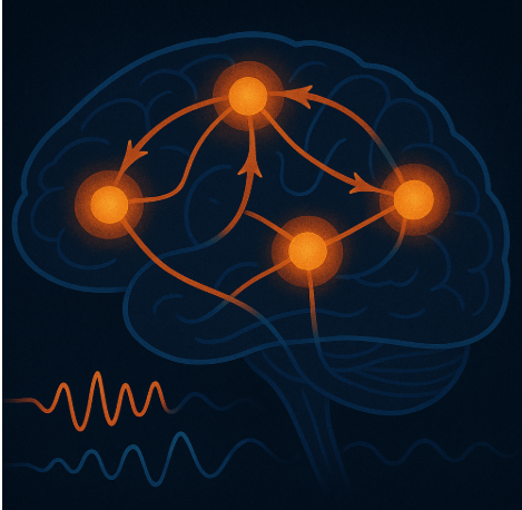
How Does Synaptic Plasticity Support Learning?
A major challenge in neuroscience is identifying the specific neurons and synapses that drive complex behaviors like reinforcement learning. To pinpoint the specific neurons and synapses that change during learning, we train mice to respond to a synthetic sensory cue—optogenetic activation of sensory cortex neurons—essentially “playing” a cue directly into the brain using light. Unlike natural sensory stimuli (e.g., sound or visual cue), this synthetic cue can be recreated in a brain slice in vitro, allowing us to measure the synaptic plasticity that accompanies the learning process using highly sensitive readouts in vitro. This novel approach bridges powerful in vivo tools (behavioral training, high-density recordings) with precise in vitro readouts (whole-cell electrophysiology, molecular profiling) to reveal how learning reshapes neural connections. In particular, we focus on how learning alters the connections from sensory cortex to the striatum, a brain region critical for decision-making and action selection. In one set of experiments, we map how learning changes the strength of these connections using optogenetics and whole-cell electrophysiology. In another set of experiments, we determine how the molecular state of the circuit evolves during learning. To do this, we combine trans-synaptic tracing with single-cell RNA sequencing. Hence, we dissect how brain circuits rewire to support learning.

How Do Distributed Brain Systems Coordinate Learning and Memory?
Learning and memory consolidation demand the coordinated interplay of multiple brain systems. The mechanisms orchestrating this cross-systems cooperation remain enigmatic. To investigate, we employ a versatile toolkit: 1) We dynamically manipulate neural activity in vivo to either pause learning or allow it to proceed. 2) We observe the effects on behavior. 3) We also observe the effects on brain activity by recording from multiple brain systems simultaneously. This approach empowers us to precisely measure the neural activity changes that accompany learning in real time, as learning pauses or it continues. By this approach, we aim to decipher the algorithms used by the distributed brain systems to learn and remember.
Might the Basal Ganglia Contribute to, or Be Able to Reverse, Tinnitus?
Tinnitus is an auditory disorder associated with anxiety, sleep disturbances, difficulty concentrating, and reduced quality of life. It is common, affecting approximately 15% of the population. It is thought to result from pathological adaptations in the central brain following damage to the auditory periphery. My lab investigates how the brain’s learning mechanisms may be hijacked to produce these maladaptive changes. One promising but understudied research direction is the role of the basal ganglia—a brain system long recognized for its critical role in plasticity and learning. Yet how auditory-basal ganglia circuits are altered in tinnitus remains unknown. Using a mouse model, we aim to induce tinnitus and measure the resulting plastic changes at the synapses between the auditory system and the basal ganglia. Ultimately, we hope to manipulate activity within these circuits to harness the brain’s natural learning machinery and reverse the pathological changes that give rise to tinnitus.

Does Learning Shift How Sensations Recruit the Striatum?
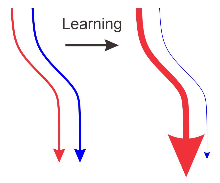
The sensory cortex sends information to a key brain region, called the striatum, that is conserved across species from fish to birds to mammals. Exiting the striatum, two main output pathways carry information to other brain regions. The balance of activity between these output pathways—much like a seesaw—is thought to either promote or suppress future actions. Hence, this balance directs learning and is important to elicit a consistent, adaptive behavioral response. Furthermore, disruptions in this balance are linked to a range of neurological and psychiatric disorders, including Huntington’s, Parkinson’s, Tourette’s, dystonia, OCD, autism, schizophrenia, addiction, and likely many others. Prior studies show that, before learning, sensory cortex inputs are evenly distributed across the two output pathways of the striatum. We hypothesize that learning shifts this balance. To test this, our lab records from specific neuron types in the two striatum pathways to track how sensory responses evolve during learning. In parallel, we use fiber photometry to measure dopamine release into the striatum, aiming to understand how dopamine shifts this balance. Our work seeks to reveal how a sensory cue, through striatum-mediated learning, comes to elicit a consistent, adaptive behavioral response.
Past Few Years
(in collaboration with the Sabatini and Scanziani labs)
What Is the Striatum’s Function in Learning and Memory Recall?
We discovered that a brain region called the posterior dorsomedial striatum tail (pDMSt) is essential for learning new actions based on sensory cues—but not for remembering them later. Like a teacher guiding early practice, the striatum helps the brain to improve with each attempt, then hands off the memory to other brain regions once the skill is learned.
Reinhold K, Iadarola M, Tang S, Chang A, Kuwamoto W, Albanese MA, Sun S, Hakim R, Zimmer J, Wang W, Sabatini BL. Striatum supports fast learning but not memory recall. Nature. 2025 May 7. doi:10.1038/s41586-025-08969-1. Epub 2025 May 7.
How Does Internal State Modulate Sensory Processing?
Animals shift between states of focused attention and sensory disengagement (“daydreaming”), but the neural mechanisms behind these shifts were not well understood. We found that feedback from the cortex to the thalamus plays a key role: it boosts thalamic sensory responses during attentive states and suppresses them during disengaged states.
Reinhold K*, Resulaj A*, Scanziani M. Brain state-dependent modulation of thalamic sensory processing by cortico-thalamic feedback. Journal of Neuroscience. 2023 Mar 1;43(9):1540-1554. PMID: 36653192; PMCID: PMC10008059; DOI: 10.1523/JNEUROSCI.2124-21.2022.